

Age Related Macular Degeneration
Dr.Maneesh Bapaye
DNB, FRCS(G), FMRF,FICO,MNAMS,MBA
Fellow-Sankara Nethralaya, Chennai
Consultant Vitreoretinal Surgeon, Dr.Bapaye Hospital, Nashik
Age-related macular degeneration (AM.D.) is a major cause of blindness in patients above the age of 55-60 years. The disease is more prevalent in western countries but is on the rise in the Indian subcontinent as well. The vision lost due to A.M.D cannot be restored. But if it is diagnosed in time, the progression of the disease can be slowed down and the existing vision can be maintained.
Structure of an Eye:
The eye works like a camera & the retina is the film of this camera. The center of the retina is the most sensitive spot of vision known as the macula. It is responsible for finalities of vision like reading, recognizing details, facial identification, etc. Diseases of the macula cause severe damage to the vision. The light rays entering the eye are focused by the cornea and the lens onto the retina.
The choroid is a layer outside the retina. It is made of densely packed blood vessels and provides blood supply to different parts of the eye. In the normal eye, it is separated from the retina by a thin layer of cells known as retinal pigment epithelium. Hence choroidal blood vessels can not leak under the retina.
The image formed on the retina is transmitted to the brain through the optic nerve. In the brain, in the visual cortex, the image is analyzed.
Symptoms of A.M.D:
- In the early stages of disease straight lines appear curved. This is known as metamorphopsia. It worsens over time if untreated.
2) While reading, the letters appear mixed with each other, some of the letters might be skipped.
3) As the disease progresses, a black spot appears in the center of the eye. This is when patients start feeling difficulty in recognizing faces, reading, and performing fine activities.
4) If untreated, with time this black spot grows in size and appears darker. In late stages day to day activities may also get affected.(Fig.1).
Types of AMD
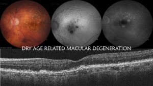
1) Dry A.M.D: This type of disease occurs in more than 80% of patients. In this type, the macula and the retina near it become thinner. Yellowish lumps called Drusen appear on the macula. As a result, the photoreceptors, cells responsible for the formation of images, in the macula are gradually destroyed. In this type, the central vision gradually decreases over a period of many months to years. But this type cannot be treated completely. The growth of the disease can be slowed down by using antioxidant drugs as well as by stopping tobacco use. (Fig.2)
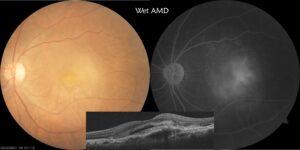
2) Wet A.M.D: This type involves the growth of choroidal blood vessels under the macula. In the early stages, the fluid component in blood seeps through these blood vessel walls and the macula becomes edematous. Over time, the blood vessels grow under the RPE cells and the retina and are known as Choroidal Neovascular Membrane ( CNVM). These may bleed under the retina. As CNVM grows in size, the symptoms mentioned above begin to increase. (Fig.3)
3) Polypoidal Choroidal Vasculopathy (PCV) is a rare type of AMD: It is more common in Asia than in the western world. This involves the formation of highly dilated blood vessels (polyps) in the area of the macula. There is a possibility of sudden massive bleeding under the retina. The treatment of this disease lasts longer and the visual recovery may be lesser.
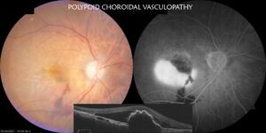
Fig.4
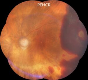
This disease can also occur in the outer part of the retina. This is called PEHCR (Fig.5)
Retinal examinations
1) Funduscopy – The retina of the eye is enlarged and examined with an indirect ophthalmoscope.
2) Fundus Photography– Fundus camera is used to take pictures of different parts of the retina and make a map of the retina. It is used to understand the nature of AMD and for future reference.
3) Fluorescein Angiography (F. F. A) – In which fluorescein dye is injected into the vein of the arm. Serial photographs are taken as it reaches retinal blood vessels. The dye does not leak through normal retinal blood vessels. However, areas of vascular abnormality can leak or block the dye which gives us an idea of disease. ( YouTube:https://youtu.be/m7_y1HnUb2Q)
4) Indocyanine Green Angiography (ICG Angiography) – Indocyanine Green is a drug that is injected intravenously and angiography of the retina and choroid is performed. This test is essential in PCV
5) Optical Coherence Tomography (OCT) – This sophisticated test can be performed in a few minutes without any injection. It accurately measures the thickness and structure of the macula. It also gives details of structures in the immediate proximity of the macula. Therefore, this test has become an essential and mainstay for the diagnosis and follow-up of AMD patients. (YouTube: https://youtu.be/pLEIO-G1K14)
Treatment methods:
1) Anti-VEGF (Fig.5) – In AMD as well as other retinal disorders, inflammatory elements in blood, VEGF increases to a large extent and adversely affects the retinal blood vessels. Drugs/injections that reduce this factor are the mainstays of AMD treatment. These injections constrict the anomalous blood vessels that have grown under the retina and reduce the chances of their regeneration and growth. At the beginning of treatment, 3 to 4 injections are given once a month known as a loading dose (Fig.6). The effect of injections usually lasts for up to 4 to 6 weeks. After the loading dose, the patients have to be examined for 4 to 8 weeks. If AMD is found to be recurring, injections have to be given. Therefore, it is impossible to predict exactly how many injections a patient will need. Avastin (Bevacizumab), Accentrix / Razumab (Ranibizumab), Pagenex (Brolicizumab), Eylea (Aflibercept) injections are currently available (Fig.7) and new injections are becoming available. The doctor decides which injection is right for which patient and suggests accordingly. (YouTube:https://youtu.be/Qce6MBuZfHI)
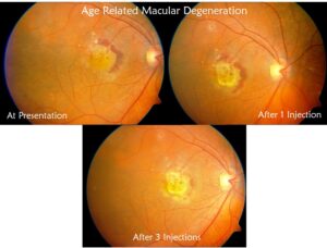
Fig.6
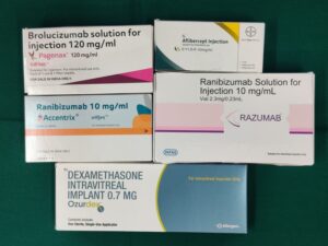
B) Steroids (Triamcinolone) and Ozurdex (Dexamethasone implant): This treatment modality is used in very few patients who have recurrent or very severe disease. May be used alone or in association with Anti VEGF injections.
2) Laser treatment – Photo Dynamic Therapy (P.D.T) or Trans Pupillary Thermotherapy (T.T.T) laser treatment is used in the disease. Of these, P.D.T. This laser system is more useful. In this laser treatment, an injection of Visudyne dye is given in the veins of the arm. The dye gets concentrated in areas of CNVM. The area of CNVM is calculated with the help of a computer. A cold laser is administered over this area which stimulates Visudyne leading to constriction and closure of CNVM
3) Combined treatment – PDT laser and one of the above injections are used together. Since these two treatment systems work differently, the combined solution is more useful than any one type.
Remember:
1) A.M.D. The disease occurs in both eyes. If the disease is in one eye, the risk of it developing in the other eye is higher. So it is essential to have regular follow-ups for another eye.
2) Amsler test (Fig.8) is given to patients at home. It should be done on a daily basis. This test is useful for monitoring the recurrence of the disease in the affected eye and early diagnosis of the disease in the unaided eye.
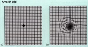
Fig. 8
3) Smoking, high blood pressure (hypertension), fast food, etc increase the risk of AMD and should be avoided. Tobacco consumption in any format must be stopped.
4) After 50 years of age, a complete eye examination should be done every year and it should be emphasized that retina examination is also done as a part of it.
(This Document is meant for patient education purposes. For Pvt Circulation Only)